Heart Valve Disease
Anatomy of the Heart 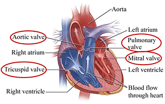
How do the heart valves function?
The heart has four valves: the aortic, tricuspid, pulmonary and mitral. These valves have tissue flaps, also known as leaflets, which open and close with each heartbeat. When the valves open blood flows through the heart chambers. When the valves close they prevent blood from flowing backwards.
When functioning properly blood flows one-way through your heart. It comes in through the top chamber (the left atrium) of the heart and passes down to the bottom chamber (the left ventricle) and to your body. As blood builds in the right and left atrium, the tricuspid and mitral valves open, then quickly shut, to allow blood to flow into the ventricles. The aortic and pulmonary valves control the blood flow out of the ventricles into your body.
What is heart valve disease?
More than 5 million Americans are diagnosed every year with heart valve disease and/or heart murmurs. Heart valve disease occurs when one or more of your heart valves aren’t functioning properly and may disrupt the way blood flows through the heart.
Valvular regurgitation or valve regurgitation and valve stenosis are the two most common heart valve problems. The most common valves affected by heart valve disease are the aortic and mitral valves. The name of the affected valve appears before the identified problem.
Depending on the severity of the valve problem, surgeons may need to repair or replace your heart valve.
Heart Center Quick Links
About Us